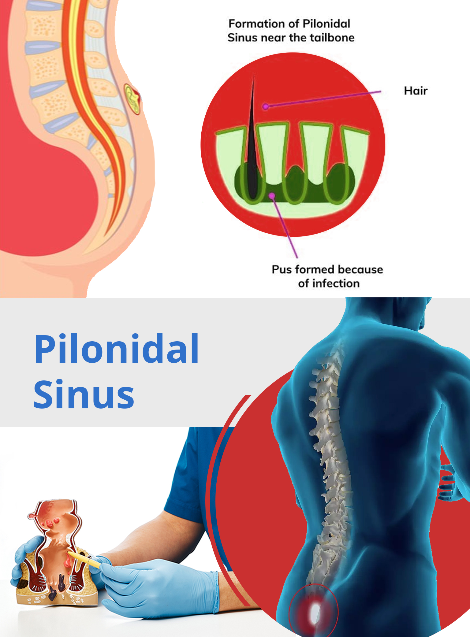
A pilonidal Sinus usually surfaces when a hair follicle in the crease of the buttocks grows inwards into the skin. The body considers this as a foreign body and launches an inflammatory response against it. This leads to the formation of an abscess which ultimately bursts. This opening in the skin continues to discharge pus and forms a sinus tract. This pus discharge becomes chronic because of the presence of the hairs acting as foreign body. The opening of the sinus discharges pus of and on and does not respond to any antibiotics. It is a painful condition and needs surgical intervention.

Anyone can get a pilonidal sinus but some people are at a higher risk than others:
What are the symptoms of an infected Pilonidal sinus?
The doctor/ surgeon can easily examine the crease of the buttocks and the sinus is visible to naked eye as a hole on the top of the cleft of the area. If it is infected, a discharge of pus/bloos is seen oozing out from the area.
The preferred method of managing this chronic condition is a minimally invasive procedure which is now the gold standard for the treatment for this modality. This modality is best suited in early cases when the skin over the sinus is healthy and is known as Endoscopic Pilonidal Sinus treatment (EPSiT).
Depending on the severity sometimes other temporary fixes are advised (like antibiotics/pain killers/ anti inflammatory medicines) however, they are only limited to providing momentary relief of symptoms in terms of pain and inflammation as these have a very high probability of recurrence.
In delayed cases the skin becomes unhealthy with multiple openings. In such cases the only alternative is Surgical excision of the sinus tract along with the bunch of hairs inside.
EPSiT is a minimally invasive technique where a fistuloscope is used to enter through the external opening. The fistuloscope has an 8 degree angle eyepiece and is equipped with an optical channel, an operative channel & an irrigation channel. As a first step which is a diagnostic step, the anatomy of the tracts and sinus cavity is visualized. This is followed by the procedure where an electrode is introduced through the operative channel and the cavity and fistula’s track are removed. All the granulation tissue is destroyed and removed. If hair is found during the procedure, they are also removed with tongs. The continuous lavage of the washing solution ensures complete elimination of debris & blood.
Usually after the procedure, the patient is walking around on the same day with minimal discomfort. They can resume their normal activities without any restrictions. The dressing on external opening is changed as required and patient is usually discharged in few hours unless required to stay for insurance purposes.